Complete Solutions and Summary of Breathing and Exchange of Gases – NCERT Class 11, Biology, Chapter 14 – Summary, Questions, Answers, Extra Questions
Summary of respiratory organs, breathing mechanism, gas exchange, transport, control, and disorders with key NCERT questions.
Updated: 8 months ago
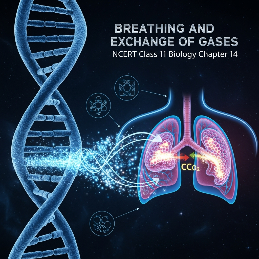
Breathing and Exchange of Gases
Chapter 14: Biology - Ultimate Study Guide | NCERT Class 11 Notes, Questions, Examples & Quiz 2025
Full Chapter Summary & Detailed Notes - Breathing and Exchange of Gases Class 11 NCERT
Overview & Key Concepts
- Chapter Goal: Understand the respiratory system, mechanisms of breathing, gas exchange, transport, regulation, and disorders. Exam Focus: Human respiratory anatomy, partial pressures, hemoglobin binding, volumes/capacities. 2025 Updates: Emphasis on pollution-related disorders, climate impact on respiration. Fun Fact: Alveoli surface area equals a tennis court (~70 m²). Core Idea: Respiration ensures O₂ supply for energy and CO₂ removal. Real-World: Asthma from urban pollution, emphysema from smoking.
- Wider Scope: Links to circulation (Ch 15), cellular respiration (Ch 12), neural control (Ch 18).
14.1 Respiratory Organs
Respiration is the exchange of O₂ from atmosphere with CO₂ from cells, essential for energy via catabolism of glucose, amino acids, fatty acids. O₂ utilization indirectly breaks down nutrients; CO₂, a harmful byproduct, must be expelled. Place hands on chest to feel breathing movement.
- Variations Across Animals: Depends on habitat/organization. Lower invertebrates (sponges, coelenterates, flatworms) use simple diffusion over body surface. Earthworms: Moist cuticle. Insects: Tracheal tubes for air transport. Aquatic arthropods/molluscs: Gills (branchial respiration). Terrestrial: Lungs (pulmonary). Vertebrates: Fishes (gills), amphibians/reptiles/birds/mammals (lungs); frogs also cutaneous via moist skin.
- Human Respiratory System (Detailed Anatomy): Paired external nostrils above upper lip lead to nasal chamber via nasal passage. Nasal chamber opens to pharynx (common food-air passage). Pharynx to larynx (sound box, cartilaginous) to trachea (straight tube to mid-thoracic cavity, divides at 5th thoracic vertebra into right/left primary bronchi). Bronchi branch into secondary/tertiary bronchi, bronchioles, terminal bronchioles. Supported by incomplete C-shaped cartilaginous rings. Terminal bronchioles form thin, irregular-walled, vascularized alveoli (bag-like). Branching network = lungs (two, covered by double-layered pleura with pleural fluid reducing friction). Outer pleura contacts thoracic lining; inner with lung surface.
- Conducting vs Respiratory Parts: External nostrils to terminal bronchioles = conducting (transports air, filters foreign particles, humidifies, warms to body temp). Alveoli/ducts = respiratory/exchange part (diffusion site). Thoracic chamber (air-tight): Vertebral column (dorsal), sternum (ventral), ribs (lateral), dome-shaped diaphragm (inferior). Volume changes in thorax reflect in lungs, essential for breathing.
- Steps of Respiration: (i) Pulmonary ventilation (breathing: in O₂, out CO₂). (ii) Diffusion O₂/CO₂ across alveolar membrane. (iii) Blood transport of gases. (iv) Diffusion between blood/tissues. (v) Cellular respiration (O₂ use, CO₂ release – Ch 12).
Figure 14.1: Diagrammatic view of human respiratory system (left lung sectional).
14.2 Mechanism of Breathing
Two stages: Inspiration (air in) and expiration (air out) via pressure gradient between lungs/atmosphere. Inspiration: Intra-pulmonary pressure < atmospheric (negative). Expiration: Intra-pulmonary > atmospheric.
- Muscles Involved: Diaphragm contracts (antero-posterior axis increase), external intercostals lift ribs/sternum (dorso-ventral increase). Overall thoracic volume up → pulmonary volume up → pressure down → air forced in. Relaxation: Diaphragm/intercostals return to normal, volume down → pressure up → air expelled. Additional abdominal muscles for forced breathing. Average: 12-16 breaths/min. Measured by spirometer for clinical assessment.
- Respiratory Volumes and Capacities (Detailed):
- Tidal Volume (TV): Normal breath air (~500 mL); 6000-8000 mL/min.
- Inspiratory Reserve Volume (IRV): Forced inspiration beyond TV (2500-3000 mL).
- Expiratory Reserve Volume (ERV): Forced expiration beyond TV (1000-1100 mL).
- Residual Volume (RV): Air left after forced expiration (1100-1200 mL).
- Capacities: Inspiratory Capacity (IC = TV + IRV). Expiratory Capacity (EC = TV + ERV). Functional Residual Capacity (FRC = ERV + RV). Vital Capacity (VC = ERV + TV + IRV). Total Lung Capacity (TLC = VC + RV).
Figures 14.2a/b: Inspiration/expiration mechanisms.
14.3 Exchange of Gases
Primary sites: Alveoli (lungs) and tissues. Simple diffusion via pressure/concentration gradient. Factors: Solubility (CO₂ 20-25x > O₂), membrane thickness (<1 mm).
- Partial Pressures: pO₂ (individual gas contribution). Table 14.1: Atmosphere (O₂ 159 mmHg, CO₂ 0.3); Alveoli (104, 40); Deoxygenated blood (40, 45); Oxygenated blood (95, 40); Tissues (40, 45). Gradients: O₂ alveoli → blood → tissues; CO₂ tissues → blood → alveoli.
- Diffusion Membrane: Three layers – alveolar squamous epithelium, capillary endothelium, basement substance. Thin for efficient diffusion. Figure 14.3: Gas exchange diagram. Figure 14.4: Alveolus-capillary section.
14.4 Transport of Gases
Blood medium: 97% O₂ by RBCs, 3% dissolved plasma. CO₂: 20-25% RBCs, 70% bicarbonate, 7% dissolved plasma.
- 14.4.1 Transport of Oxygen: Hemoglobin (Fe-pigment in RBCs) binds reversibly to oxyhemoglobin (4 O₂ max/molecule). Binding: pO₂ primary; affected by pCO₂, H⁺, temperature. Sigmoid oxygen dissociation curve (Fig 14.5): High pO₂/low pCO₂/low H⁺/low temp in alveoli → binding; opposite in tissues → dissociation. 100 mL oxygenated blood delivers ~5 mL O₂.
- 14.4.2 Transport of Carbon Dioxide: 20-25% carbaminohemoglobin (pCO₂-related; low pO₂ aids binding in tissues, high pO₂ aids release in alveoli). 70% bicarbonate via carbonic anhydrase: CO₂ + H₂O ⇌ H₂CO₃ ⇌ H⁺ + HCO₃⁻ (RBCs/plasma). High pCO₂ in tissues → bicarbonate formation; low in alveoli → reverse. 100 mL deoxygenated blood delivers ~4 mL CO₂.
14.5 Regulation of Respiration
Neural control maintains rhythm per tissue demands. Respiratory rhythm center (medulla) primary; pneumotaxic center (pons) moderates inspiration duration/rate. Chemosensitive area (medulla) sensitive to CO₂/H⁺ → signals rhythm center. Aortic/carotid receptors detect CO₂/H⁺ changes → signals. O₂ role minor.
14.6 Disorders of Respiratory System
- Asthma: Wheezing from bronchi/bronchioles inflammation/narrowing.
- Emphysema: Chronic; alveolar walls damaged → reduced surface. Major cause: Smoking.
- Occupational: Dust in grinding/stone-breaking → inflammation/fibrosis/lung damage. Use masks.
Summary
Cells use O₂ for metabolism, produce CO₂. Evolved mechanisms for transport/removal. Human system: Lungs/air passages. Steps: Ventilation, diffusion, transport, tissue exchange, cellular respiration. Volumes/capacities via spirometer. Diffusion via gradients/solubility/thinness. O₂ as oxyhemoglobin; CO₂ as bicarbonate/carbamino. Rhythm: Medulla/pneumotaxic/chemosensitive centers.
Why This Guide Stands Out
Complete coverage: All subtopics, diagrams explained, Q&A (NCERT + extras), quiz. Exam-ready for 2025. Free & ad-free.
Key Themes & Tips
- Respiration Essentials: O₂ in, CO₂ out for energy.
- Human Focus: Anatomy, volumes, transport curves.
- Tip: Memorize partial pressures table; draw dissociation curve.
Exam Case Studies
Questions on volumes (e.g., VC calculation), disorders (smoking effects).
Project & Group Ideas
- Measure TV via balloon; discuss pollution impact on lungs.


As an Amazon Associate, ProSyllabus earns from qualifying purchases. Prices shown are subject to change.
Test your CBSE Class 11 Annual Assessment prep
Quizzes
10 questions · ~10 minutes · instant rank & AI diagnosis
Accountancy (Class 11) Practice Quiz | CBSE Class 11 Annual Assessment
Sets and Venn Operations Fundamentals — Free CBSE Class 11 Annual Assessment Quiz
Class 11 English — The Tale of Melon City (Practice Quiz)
Class 11 English — Birth (Practice Quiz)
Class 11 English — Mother's Day (Practice Quiz)
Class 11 English — The Address (Practice Quiz)
Class 11 English — The Summer of the Beautiful White Horse (Practice Quiz)
Class 11 English — Father to Son (Practice Quiz)
Class 11 English — Silk Road (Practice Quiz)
Class 11 English — The Adventure (Practice Quiz)
Class 11 English — Childhood (Practice Quiz)
Class 11 English — The Ailing Planet: the Green Movement's Role (Practice Quiz)
Class 11 English — The Voice of the Rain (Practice Quiz)
Class 11 English — The Laburnum Top (Practice Quiz)
Class 11 English — Discovering Tut: the Saga Continues (Practice Quiz)
Class 11 English — We're Not Afraid to Die... if We Can All Be Together (Practice Quiz)
Class 11 English — A Photograph (Practice Quiz)
Class 11 English — The Portrait of a Lady (Practice Quiz)
Class 11 Psychology — Motivation and Emotion (Practice Quiz)
Class 11 Psychology — Thinking (Practice Quiz)
Class 11 Psychology — Human Memory (Practice Quiz)
Class 11 Psychology — Learning (Practice Quiz)
Class 11 Psychology — Sensory, Attentional and Perceptual Processes (Practice Quiz)
Class 11 Psychology — Human Development (Practice Quiz)
Class 11 Psychology — Methods of Enquiry in Psychology (Practice Quiz)
Class 11 Psychology — What is Psychology? (Practice Quiz)
Class 11 Sociology — Indian Sociologists (Practice Quiz)
Class 11 Sociology — Introducing Western Sociologists (Practice Quiz)
Class 11 Sociology — Environment and Society (Practice Quiz)
Class 11 Sociology — Social Change and Social Order in Rural and Urban Society (Practice Quiz)
Class 11 Sociology — Social Structure, Stratification and Social Processes in Society (Practice Quiz)
Class 11 Sociology — Doing Sociology: Research Methods (Practice Quiz)
Class 11 Sociology — Culture and Socialisation (Practice Quiz)
Class 11 Sociology — Understanding Social Institutions (Practice Quiz)
Class 11 Sociology — Terms, Concepts and Their Use in Sociology (Practice Quiz)
Class 11 Sociology — Sociology and Society (Practice Quiz)
Class 11 Political Science — The Philosophy of the Constitution (Practice Quiz)
Class 11 Political Science — Constitution as a Living Document (Practice Quiz)
Class 11 Political Science — Local Governments (Practice Quiz)
Class 11 Political Science — Federalism (Practice Quiz)
Class 11 Political Science — Judiciary (Practice Quiz)
Class 11 Political Science — Legislature (Practice Quiz)
Class 11 Political Science — Executive (Practice Quiz)
Class 11 Political Science — Election and Representation (Practice Quiz)
Class 11 Political Science — Rights in the Indian Constitution (Practice Quiz)
Class 11 Political Science — Constitution: Why and How? (Practice Quiz)
Class 11 Political Science — Secularism (Practice Quiz)
Class 11 Political Science — Nationalism (Practice Quiz)
Class 11 Political Science — Citizenship (Practice Quiz)
Class 11 Political Science — Rights (Practice Quiz)
Class 11 Political Science — Social Justice (Practice Quiz)
Class 11 Political Science — Equality (Practice Quiz)
Class 11 Political Science — Freedom (Practice Quiz)
Class 11 Political Science — Political Theory: An Introduction (Practice Quiz)
Class 11 Geography — Natural Hazards and Disasters (Practice Quiz)
Class 11 Geography — Natural Vegetation (Practice Quiz)
Class 11 Geography — Climate (Practice Quiz)
Class 11 Geography — Drainage System (Practice Quiz)
Class 11 Geography — Structure and Physiography (Practice Quiz)
Class 11 Geography — India – Location (Practice Quiz)
Class 11 Geography — Biodiversity and Conservation (Practice Quiz)
Class 11 Geography — Movements of Ocean Water (Practice Quiz)
Class 11 Geography — Water (Oceans) (Practice Quiz)
Class 11 Geography — World Climate and Climate Change (Practice Quiz)
Class 11 Geography — Water in the Atmosphere (Practice Quiz)
Class 11 Geography — Atmospheric Circulation and Weather Systems (Practice Quiz)
Class 11 Geography — Solar Radiation, Heat Balance and Temperature (Practice Quiz)
Class 11 Geography — Composition and Structure of Atmosphere (Practice Quiz)
Class 11 Geography — Landforms and their Evolution (Practice Quiz)
Class 11 Geography — Geomorphic Processes (Practice Quiz)
Class 11 Geography — Distribution of Oceans and Continents (Practice Quiz)
Class 11 Geography — Interior of the Earth (Practice Quiz)
Class 11 Geography — The Origin and Evolution of the Earth (Practice Quiz)
Class 11 Geography — Geography as a Discipline (Practice Quiz)
Class 11 History — Paths to Modernisation (Practice Quiz)
Class 11 History — Displacing Indigenous Peoples (Practice Quiz)
Class 11 History — Changing Cultural Traditions (Practice Quiz)
Class 11 History — The Three Orders (Practice Quiz)
Class 11 History — Nomadic Empires (Practice Quiz)
Class 11 History — An Empire Across Three Continents (Practice Quiz)
Class 11 History — Writing and City Life (Practice Quiz)
Class 11 Economics — Comparative Development Experiences of India and its Neighbours (Practice Quiz)
Class 11 Economics — Environment and Sustainable Development (Practice Quiz)
Class 11 Economics — Employment: Growth, Informalisation and Other Issues (Practice Quiz)
Class 11 Economics — Rural Development (Practice Quiz)
Class 11 Economics — Human Capital Formation in India (Practice Quiz)
Class 11 Economics — Liberalisation, Privatisation and Globalisation: An Appraisal (Practice Quiz)
Class 11 Economics — Indian Economy 1950-1990 (Practice Quiz)
Class 11 Economics — Indian Economy on the Eve of Independence (Practice Quiz)
Class 11 Economics — Use of Statistical Tools (Practice Quiz)
Class 11 Economics — Index Numbers (Practice Quiz)
Class 11 Economics — Correlation (Practice Quiz)
Class 11 Economics — Measures of Central Tendency (Practice Quiz)
Class 11 Economics — Presentation of Data (Practice Quiz)
Class 11 Economics — Organisation of Data (Practice Quiz)
Class 11 Economics — Collection of Data (Practice Quiz)
Class 11 Economics — Introduction (Practice Quiz)
Class 11 Business Studies — International Business (Practice Quiz)
Class 11 Business Studies — Internal Trade (Practice Quiz)
Class 11 Business Studies — MSME and Business Entrepreneurship (Practice Quiz)
Class 11 Business Studies — Sources of Business Finance (Practice Quiz)
Class 11 Business Studies — Formation of a Company (Practice Quiz)
Class 11 Business Studies — Social Responsibilities of Business and Business Ethics (Practice Quiz)
Class 11 Business Studies — Emerging Modes of Business (Practice Quiz)
Class 11 Business Studies — Business Services (Practice Quiz)
Class 11 Business Studies — Private, Public and Global Enterprises (Practice Quiz)
Class 11 Business Studies — Forms of Business Organisation (Practice Quiz)
Class 11 Business Studies — Business, Trade and Commerce (Practice Quiz)
Class 11 Accountancy — Financial Statements - II (Practice Quiz)
Class 11 Accountancy — Financial Statements - I (Practice Quiz)
Class 11 Accountancy — Depreciation, Provisions and Reserves (Practice Quiz)
Class 11 Accountancy — Trial Balance and Rectification of Errors (Practice Quiz)
Class 11 Accountancy — Bank Reconciliation Statement (Practice Quiz)
Class 11 Accountancy — Recording of Transactions - II (Practice Quiz)
Class 11 Accountancy — Recording of Transactions - I (Practice Quiz)
Class 11 Accountancy — Theory Base of Accounting (Practice Quiz)
Class 11 Accountancy — Introduction to Accounting (Practice Quiz)
Class 11 Maths — Probability (Practice Quiz)
Class 11 Maths — Statistics (Practice Quiz)
Class 11 Maths — Limits and Derivatives (Practice Quiz)
Class 11 Maths — Introduction to Three Dimensional Geometry (Practice Quiz)
Class 11 Maths — Conic Sections (Practice Quiz)
Class 11 Maths — Straight Lines (Practice Quiz)
Class 11 Maths — Sequences and Series (Practice Quiz)
Class 11 Maths — Binomial Theorem (Practice Quiz)
Class 11 Maths — Permutations and Combinations (Practice Quiz)
Class 11 Maths — Linear Inequalities (Practice Quiz)
Class 11 Maths — Complex Numbers and Quadratic Equations (Practice Quiz)
Class 11 Maths — Relations and Functions (Practice Quiz)
Class 11 Maths — Sets (Practice Quiz)
Class 11 Biology — Chemical Coordination and Integration (Practice Quiz)
Class 11 Biology — Neural Control and Coordination (Practice Quiz)
Class 11 Biology — Locomotion and Movement (Practice Quiz)
Class 11 Biology — Excretory Products and their Elimination (Practice Quiz)
Class 11 Biology — Body Fluids and Circulation (Practice Quiz)
Class 11 Biology — Breathing and Exchange of Gases (Practice Quiz)
Class 11 Biology — Plant Growth and Development (Practice Quiz)
Class 11 Biology — Respiration in Plants (Practice Quiz)
Class 11 Biology — Photosynthesis in Higher Plants (Practice Quiz)
Class 11 Biology — Cell Cycle and Cell Division (Practice Quiz)
Class 11 Biology — Biomolecules (Practice Quiz)
Class 11 Biology — Cell: The Unit of Life (Practice Quiz)
Class 11 Biology — Structural Organisation in Animals (Practice Quiz)
Class 11 Biology — Anatomy of Flowering Plants (Practice Quiz)
Class 11 Biology — Morphology of Flowering Plants (Practice Quiz)
Class 11 Biology — Animal Kingdom (Practice Quiz)
Class 11 Biology — Plant Kingdom (Practice Quiz)
Class 11 Biology — Biological Classification (Practice Quiz)
Class 11 Biology — The Living World (Practice Quiz)
Class 11 Chemistry — Hydrocarbons (Practice Quiz)
Class 11 Chemistry — Organic Chemistry – Some Basic Principles and Techniques (Practice Quiz)
Class 11 Chemistry — Redox Reactions (Practice Quiz)
Class 11 Chemistry — Equilibrium (Practice Quiz)
Class 11 Chemistry — Chemical Bonding and Molecular Structure (Practice Quiz)
Class 11 Chemistry — Classification of Elements and Periodicity in Properties (Practice Quiz)
Class 11 Chemistry — Some Basic Concepts of Chemistry (Practice Quiz)
Class 11 Physics — Waves (Practice Quiz)
Class 11 Physics — Oscillations (Practice Quiz)
Class 11 Physics — Kinetic Theory (Practice Quiz)
Class 11 Chemistry — Thermodynamics (Practice Quiz)
Class 11 Physics — Thermal Properties of Matter (Practice Quiz)
Class 11 Physics — Mechanical Properties of Fluids (Practice Quiz)
Class 11 Physics — Mechanical Properties of Solids (Practice Quiz)
Class 11 Physics — Gravitation (Practice Quiz)
Class 11 Physics — Systems of Particles and Rotational Motion (Practice Quiz)
Class 11 Physics — Work, Energy and Power (Practice Quiz)
Class 11 Physics — Motion in a Plane (Practice Quiz)
Class 11 Physics — Motion in a Straight Line (Practice Quiz)
Class 11 Physics — Units and Measurement (Practice Quiz)
Class 11 Maths — Trigonometric Functions (Practice Quiz)
Class 11 Chemistry — Structure of Atom (Practice Quiz)
Class 11 Physics — Laws of Motion (Practice Quiz)
Business Studies (Class 11) Practice Quiz | CBSE Class 11 Annual Assessment
Economics (Class 11) Practice Quiz | CBSE Class 11 Annual Assessment
Humanities Subjects Practice Quiz | CBSE Class 11 Annual Assessment
Motion in a Straight Line Practice Quiz | CBSE Class 11 Annual Assessment
Thermodynamic Processes and Laws Advanced Challenge | CBSE Class 11 Annual Assessment

Group Discussions
No forum posts available.

