Complete Solutions and Summary of Locomotion and Movement – NCERT Class 11, Biology, Chapter 17 – Summary, Questions, Answers, Extra Questions
Summary of types of movement, skeletal system, muscles, mechanism of locomotion in animals, human skeleton, joints, and muscular activities with important NCERT exercises.
Updated: 8 months ago
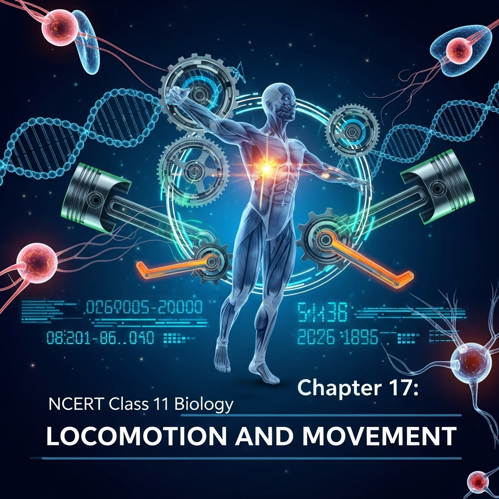
Locomotion and Movement
Chapter 17: Biology - Ultimate Study Guide | NCERT Class 11 Notes, Questions, Examples & Quiz 2025
Full Chapter Summary & Detailed Notes - Locomotion and Movement Class 11 NCERT
Overview & Key Concepts
- Chapter Goal: Explore types of movements in living organisms, focusing on human locomotion involving muscular, skeletal, and neural systems. Exam Focus: Muscle types, sarcomere structure, sliding filament theory, skeletal components, joints, disorders. 2025 Updates: Emphasis on molecular mechanisms and health implications. Fun Fact: Human muscles make up 40-50% of body weight, enabling precise control. Core Idea: Movements range from protoplasmic streaming to complex locomotion, all linked to survival needs like food search and predator escape. Real-World: Understanding muscle contraction aids in sports science and rehabilitation.
- Wider Scope: Links to physiology, evolution (e.g., limb adaptations), and medicine (e.g., myopathies).
Introduction: Movement and Locomotion
- Movement is a defining feature of life, seen in protoplasmic streaming in Amoeba, ciliary/flagellar motion in unicellular organisms, and limb/tentacle movements in multicellular ones. In humans, it includes jaw, eyelid, and tongue actions.
- Locomotion is voluntary movement changing location (e.g., walking, swimming), distinct from but linked to other movements. Structures like cilia in Paramoecium serve dual roles in feeding and movement; Hydra tentacles capture prey and aid locomotion.
- All locomotions are movements, but not vice versa. Animals locomote for food, shelter, mating, breeding, climate, or escape. Habitats influence methods (e.g., flying in birds, swimming in fish).
- Human locomotion requires coordinated muscular, skeletal, and neural activity, forming the chapter's core.
17.1 Types of Movement
- Human cells show three types: Amoeboid (pseudopodia-based, by macrophages/leucocytes via protoplasm streaming and microfilaments; e.g., immune response), Ciliary (coordinated cilia in tubular organs like trachea for dust removal or oviduct for egg transport), and Muscular (contractile for limb/jaw movement; basis for locomotion).
- Amoeboid: Involves cytoskeletal elements; crucial for phagocytosis. Ciliary: Lined by ciliated epithelium; prevents respiratory issues. Muscular: Detailed in later sections; enables precise control.
- These movements ensure internal transport (e.g., food/gametes) and external locomotion, highlighting integration.
17.2 Muscle: Types, Structure, and Properties
- Muscles (mesodermal, 40-50% body weight) have excitability, contractility, extensibility, elasticity. Classified by location (skeletal/voluntary/striated, visceral/involuntary/smooth, cardiac/involuntary/striated/branched), appearance, and regulation.
- Skeletal: Attached to bones, striped, voluntary; for posture/locomotion. Visceral: In organ walls (e.g., gut), smooth, involuntary; aids peristalsis/gamete transport. Cardiac: Heart-specific, striated, involuntary; self-excitable via nervous branching.
- Structure: Muscle organized in fascicles (bundles) held by fascia; each fiber (syncytium with sarcolemma, sarcoplasm, sarcoplasmic reticulum for Ca++ storage) has myofibrils with sarcomeres (functional unit between Z-lines).
- Sarcomere: Alternating A-band (thick myosin) and I-band (thin actin); H-zone (central myosin not overlapped); M-line holds thick filaments; Z-line anchors thin ones. Resting: Partial overlap, H-zone visible.
- Contractile Proteins: Actin (thin: F-actin helix with G-actin monomers, tropomyosin, troponin masking sites); Myosin (thick: Meromyosins – HMM head with ATPase/ATP-actin sites, LMM tail).
17.2.2 Mechanism of Muscle Contraction
- Sliding Filament Theory: Thin filaments slide over thick via cross-bridges, shortening sarcomere (I-band/H-zone reduce, A-band constant).
- Steps: Neural signal via motor neuron to neuromuscular junction releases acetylcholine, generating action potential; spreads, releases Ca++ from sarcoplasmic reticulum. Ca++ binds troponin, exposes actin sites. Myosin head (ATP-hydrolyzed) binds actin (cross-bridge), pulls (power stroke via ADP+Pi release), shortens sarcomere.
- Relaxation: New ATP breaks cross-bridge; Ca++ pumped back, troponin masks sites; Z-lines return. Cycle repeats for sustained contraction. Fatigue from lactic acid (anaerobic glycolysis).
- Muscle Fibers: Red (high myoglobin/mitochondria, aerobic, fatigue-resistant); White (low myoglobin, high SR, anaerobic, fast but fatigable).
17.3 Skeletal System
- Framework of 206 bones + cartilages (hard matrix with Ca salts; pliable with chondroitin). Divisions: Axial (80 bones: skull 22+hyoid+ear ossicles, vertebral column 26, sternum+ribs 25); Appendicular (limbs 60x2 + girdles 10).
- Axial Details: Skull (cranium 8 for brain protection, facial 14; dicondylic with occipital condyles; ear ossicles: malleus/incus/stapes). Vertebral Column (cervical 7, thoracic 12, lumbar 5, sacral 1 fused, coccygeal 1 fused; protects spinal cord, supports head/ribs). Rib Cage (12 bicephalic pairs: 7 true, 3 false vertebrochondral, 2 floating; with sternum for thoracic protection).
- Appendicular: Pectoral Girdle (clavicle+scapula x2; glenoid cavity for humerus). Upper Limb (humerus, radius/ulna, 8 carpals, 5 metacarpals, 14 phalanges). Pelvic Girdle (ilium/ischium/pubis fused into coxal x2; acetabulum for femur, pubic symphysis). Lower Limb (femur longest, tibia/fibula, 7 tarsals, 5 metatarsals, 14 phalanges, patella kneecap).
17.4 Joints
- Points of contact enabling movement via muscle force (fulcrum). Types: Fibrous (immovable, e.g., skull sutures), Cartilaginous (slightly movable, e.g., intervertebral), Synovial (freely movable with fluid cavity; subtypes: ball-socket humerus/pectoral, hinge knee, pivot atlas/axis, gliding carpals, saddle thumb carpal/metacarpal).
- Synovial joints crucial for locomotion; vary by movability factors like structure/ligaments.
17.5 Disorders of Muscular and Skeletal System
- Myasthenia Gravis: Autoimmune neuromuscular junction attack causing fatigue/paralysis. Muscular Dystrophy: Genetic progressive skeletal muscle degeneration. Tetany: Low Ca++-induced spasms. Arthritis: Joint inflammation. Osteoporosis: Age-related bone mass loss/fracture risk (estrogen deficiency). Gout: Uric acid crystal joint inflammation.
- These highlight need for balanced Ca++, estrogen, and genetic health.
Summary
- Movements essential for life; types amoeboid/ciliary/muscular. Muscles: Three types with unique structures/properties. Contraction via sliding filaments/Ca++ cross-bridges. Skeleton: Axial/appendicular for support/protection. Joints enable motion. Disorders affect mobility.
Why This Guide Stands Out
Complete coverage: Detailed subtopics, diagrams explained, Q&A aligned to marks, quiz for practice. Exam-ready for 2025, free & ad-free.
Key Themes & Tips
- Integration: Muscular-skeletal-neural synergy for locomotion.
- Molecular Basis: Actin-myosin-Ca++ in contraction.
- Tip: Draw sarcomere diagrams; memorize bone counts/joint types.
Exam Case Studies
Questions on sliding theory steps, axial vs. appendicular, disorder causes.
Project & Group Ideas
- Model sarcomere with clay; discuss athlete muscle types (red/white).


As an Amazon Associate, ProSyllabus earns from qualifying purchases. Prices shown are subject to change.
Test your CBSE Class 11 Annual Assessment prep
Quizzes
10 questions · ~10 minutes · instant rank & AI diagnosis
Accountancy (Class 11) Practice Quiz | CBSE Class 11 Annual Assessment
Sets and Venn Operations Fundamentals — Free CBSE Class 11 Annual Assessment Quiz
Class 11 English — The Tale of Melon City (Practice Quiz)
Class 11 English — Birth (Practice Quiz)
Class 11 English — Mother's Day (Practice Quiz)
Class 11 English — The Address (Practice Quiz)
Class 11 English — The Summer of the Beautiful White Horse (Practice Quiz)
Class 11 English — Father to Son (Practice Quiz)
Class 11 English — Silk Road (Practice Quiz)
Class 11 English — The Adventure (Practice Quiz)
Class 11 English — Childhood (Practice Quiz)
Class 11 English — The Ailing Planet: the Green Movement's Role (Practice Quiz)
Class 11 English — The Voice of the Rain (Practice Quiz)
Class 11 English — The Laburnum Top (Practice Quiz)
Class 11 English — Discovering Tut: the Saga Continues (Practice Quiz)
Class 11 English — We're Not Afraid to Die... if We Can All Be Together (Practice Quiz)
Class 11 English — A Photograph (Practice Quiz)
Class 11 English — The Portrait of a Lady (Practice Quiz)
Class 11 Psychology — Motivation and Emotion (Practice Quiz)
Class 11 Psychology — Thinking (Practice Quiz)
Class 11 Psychology — Human Memory (Practice Quiz)
Class 11 Psychology — Learning (Practice Quiz)
Class 11 Psychology — Sensory, Attentional and Perceptual Processes (Practice Quiz)
Class 11 Psychology — Human Development (Practice Quiz)
Class 11 Psychology — Methods of Enquiry in Psychology (Practice Quiz)
Class 11 Psychology — What is Psychology? (Practice Quiz)
Class 11 Sociology — Indian Sociologists (Practice Quiz)
Class 11 Sociology — Introducing Western Sociologists (Practice Quiz)
Class 11 Sociology — Environment and Society (Practice Quiz)
Class 11 Sociology — Social Change and Social Order in Rural and Urban Society (Practice Quiz)
Class 11 Sociology — Social Structure, Stratification and Social Processes in Society (Practice Quiz)
Class 11 Sociology — Doing Sociology: Research Methods (Practice Quiz)
Class 11 Sociology — Culture and Socialisation (Practice Quiz)
Class 11 Sociology — Understanding Social Institutions (Practice Quiz)
Class 11 Sociology — Terms, Concepts and Their Use in Sociology (Practice Quiz)
Class 11 Sociology — Sociology and Society (Practice Quiz)
Class 11 Political Science — The Philosophy of the Constitution (Practice Quiz)
Class 11 Political Science — Constitution as a Living Document (Practice Quiz)
Class 11 Political Science — Local Governments (Practice Quiz)
Class 11 Political Science — Federalism (Practice Quiz)
Class 11 Political Science — Judiciary (Practice Quiz)
Class 11 Political Science — Legislature (Practice Quiz)
Class 11 Political Science — Executive (Practice Quiz)
Class 11 Political Science — Election and Representation (Practice Quiz)
Class 11 Political Science — Rights in the Indian Constitution (Practice Quiz)
Class 11 Political Science — Constitution: Why and How? (Practice Quiz)
Class 11 Political Science — Secularism (Practice Quiz)
Class 11 Political Science — Nationalism (Practice Quiz)
Class 11 Political Science — Citizenship (Practice Quiz)
Class 11 Political Science — Rights (Practice Quiz)
Class 11 Political Science — Social Justice (Practice Quiz)
Class 11 Political Science — Equality (Practice Quiz)
Class 11 Political Science — Freedom (Practice Quiz)
Class 11 Political Science — Political Theory: An Introduction (Practice Quiz)
Class 11 Geography — Natural Hazards and Disasters (Practice Quiz)
Class 11 Geography — Natural Vegetation (Practice Quiz)
Class 11 Geography — Climate (Practice Quiz)
Class 11 Geography — Drainage System (Practice Quiz)
Class 11 Geography — Structure and Physiography (Practice Quiz)
Class 11 Geography — India – Location (Practice Quiz)
Class 11 Geography — Biodiversity and Conservation (Practice Quiz)
Class 11 Geography — Movements of Ocean Water (Practice Quiz)
Class 11 Geography — Water (Oceans) (Practice Quiz)
Class 11 Geography — World Climate and Climate Change (Practice Quiz)
Class 11 Geography — Water in the Atmosphere (Practice Quiz)
Class 11 Geography — Atmospheric Circulation and Weather Systems (Practice Quiz)
Class 11 Geography — Solar Radiation, Heat Balance and Temperature (Practice Quiz)
Class 11 Geography — Composition and Structure of Atmosphere (Practice Quiz)
Class 11 Geography — Landforms and their Evolution (Practice Quiz)
Class 11 Geography — Geomorphic Processes (Practice Quiz)
Class 11 Geography — Distribution of Oceans and Continents (Practice Quiz)
Class 11 Geography — Interior of the Earth (Practice Quiz)
Class 11 Geography — The Origin and Evolution of the Earth (Practice Quiz)
Class 11 Geography — Geography as a Discipline (Practice Quiz)
Class 11 History — Paths to Modernisation (Practice Quiz)
Class 11 History — Displacing Indigenous Peoples (Practice Quiz)
Class 11 History — Changing Cultural Traditions (Practice Quiz)
Class 11 History — The Three Orders (Practice Quiz)
Class 11 History — Nomadic Empires (Practice Quiz)
Class 11 History — An Empire Across Three Continents (Practice Quiz)
Class 11 History — Writing and City Life (Practice Quiz)
Class 11 Economics — Comparative Development Experiences of India and its Neighbours (Practice Quiz)
Class 11 Economics — Environment and Sustainable Development (Practice Quiz)
Class 11 Economics — Employment: Growth, Informalisation and Other Issues (Practice Quiz)
Class 11 Economics — Rural Development (Practice Quiz)
Class 11 Economics — Human Capital Formation in India (Practice Quiz)
Class 11 Economics — Liberalisation, Privatisation and Globalisation: An Appraisal (Practice Quiz)
Class 11 Economics — Indian Economy 1950-1990 (Practice Quiz)
Class 11 Economics — Indian Economy on the Eve of Independence (Practice Quiz)
Class 11 Economics — Use of Statistical Tools (Practice Quiz)
Class 11 Economics — Index Numbers (Practice Quiz)
Class 11 Economics — Correlation (Practice Quiz)
Class 11 Economics — Measures of Central Tendency (Practice Quiz)
Class 11 Economics — Presentation of Data (Practice Quiz)
Class 11 Economics — Organisation of Data (Practice Quiz)
Class 11 Economics — Collection of Data (Practice Quiz)
Class 11 Economics — Introduction (Practice Quiz)
Class 11 Business Studies — International Business (Practice Quiz)
Class 11 Business Studies — Internal Trade (Practice Quiz)
Class 11 Business Studies — MSME and Business Entrepreneurship (Practice Quiz)
Class 11 Business Studies — Sources of Business Finance (Practice Quiz)
Class 11 Business Studies — Formation of a Company (Practice Quiz)
Class 11 Business Studies — Social Responsibilities of Business and Business Ethics (Practice Quiz)
Class 11 Business Studies — Emerging Modes of Business (Practice Quiz)
Class 11 Business Studies — Business Services (Practice Quiz)
Class 11 Business Studies — Private, Public and Global Enterprises (Practice Quiz)
Class 11 Business Studies — Forms of Business Organisation (Practice Quiz)
Class 11 Business Studies — Business, Trade and Commerce (Practice Quiz)
Class 11 Accountancy — Financial Statements - II (Practice Quiz)
Class 11 Accountancy — Financial Statements - I (Practice Quiz)
Class 11 Accountancy — Depreciation, Provisions and Reserves (Practice Quiz)
Class 11 Accountancy — Trial Balance and Rectification of Errors (Practice Quiz)
Class 11 Accountancy — Bank Reconciliation Statement (Practice Quiz)
Class 11 Accountancy — Recording of Transactions - II (Practice Quiz)
Class 11 Accountancy — Recording of Transactions - I (Practice Quiz)
Class 11 Accountancy — Theory Base of Accounting (Practice Quiz)
Class 11 Accountancy — Introduction to Accounting (Practice Quiz)
Class 11 Maths — Probability (Practice Quiz)
Class 11 Maths — Statistics (Practice Quiz)
Class 11 Maths — Limits and Derivatives (Practice Quiz)
Class 11 Maths — Introduction to Three Dimensional Geometry (Practice Quiz)
Class 11 Maths — Conic Sections (Practice Quiz)
Class 11 Maths — Straight Lines (Practice Quiz)
Class 11 Maths — Sequences and Series (Practice Quiz)
Class 11 Maths — Binomial Theorem (Practice Quiz)
Class 11 Maths — Permutations and Combinations (Practice Quiz)
Class 11 Maths — Linear Inequalities (Practice Quiz)
Class 11 Maths — Complex Numbers and Quadratic Equations (Practice Quiz)
Class 11 Maths — Relations and Functions (Practice Quiz)
Class 11 Maths — Sets (Practice Quiz)
Class 11 Biology — Chemical Coordination and Integration (Practice Quiz)
Class 11 Biology — Neural Control and Coordination (Practice Quiz)
Class 11 Biology — Locomotion and Movement (Practice Quiz)
Class 11 Biology — Excretory Products and their Elimination (Practice Quiz)
Class 11 Biology — Body Fluids and Circulation (Practice Quiz)
Class 11 Biology — Breathing and Exchange of Gases (Practice Quiz)
Class 11 Biology — Plant Growth and Development (Practice Quiz)
Class 11 Biology — Respiration in Plants (Practice Quiz)
Class 11 Biology — Photosynthesis in Higher Plants (Practice Quiz)
Class 11 Biology — Cell Cycle and Cell Division (Practice Quiz)
Class 11 Biology — Biomolecules (Practice Quiz)
Class 11 Biology — Cell: The Unit of Life (Practice Quiz)
Class 11 Biology — Structural Organisation in Animals (Practice Quiz)
Class 11 Biology — Anatomy of Flowering Plants (Practice Quiz)
Class 11 Biology — Morphology of Flowering Plants (Practice Quiz)
Class 11 Biology — Animal Kingdom (Practice Quiz)
Class 11 Biology — Plant Kingdom (Practice Quiz)
Class 11 Biology — Biological Classification (Practice Quiz)
Class 11 Biology — The Living World (Practice Quiz)
Class 11 Chemistry — Hydrocarbons (Practice Quiz)
Class 11 Chemistry — Organic Chemistry – Some Basic Principles and Techniques (Practice Quiz)
Class 11 Chemistry — Redox Reactions (Practice Quiz)
Class 11 Chemistry — Equilibrium (Practice Quiz)
Class 11 Chemistry — Chemical Bonding and Molecular Structure (Practice Quiz)
Class 11 Chemistry — Classification of Elements and Periodicity in Properties (Practice Quiz)
Class 11 Chemistry — Some Basic Concepts of Chemistry (Practice Quiz)
Class 11 Physics — Waves (Practice Quiz)
Class 11 Physics — Oscillations (Practice Quiz)
Class 11 Physics — Kinetic Theory (Practice Quiz)
Class 11 Chemistry — Thermodynamics (Practice Quiz)
Class 11 Physics — Thermal Properties of Matter (Practice Quiz)
Class 11 Physics — Mechanical Properties of Fluids (Practice Quiz)
Class 11 Physics — Mechanical Properties of Solids (Practice Quiz)
Class 11 Physics — Gravitation (Practice Quiz)
Class 11 Physics — Systems of Particles and Rotational Motion (Practice Quiz)
Class 11 Physics — Work, Energy and Power (Practice Quiz)
Class 11 Physics — Motion in a Plane (Practice Quiz)
Class 11 Physics — Motion in a Straight Line (Practice Quiz)
Class 11 Physics — Units and Measurement (Practice Quiz)
Class 11 Maths — Trigonometric Functions (Practice Quiz)
Class 11 Chemistry — Structure of Atom (Practice Quiz)
Class 11 Physics — Laws of Motion (Practice Quiz)
Business Studies (Class 11) Practice Quiz | CBSE Class 11 Annual Assessment
Economics (Class 11) Practice Quiz | CBSE Class 11 Annual Assessment
Humanities Subjects Practice Quiz | CBSE Class 11 Annual Assessment
Motion in a Straight Line Practice Quiz | CBSE Class 11 Annual Assessment
Thermodynamic Processes and Laws Advanced Challenge | CBSE Class 11 Annual Assessment

Group Discussions
No forum posts available.

